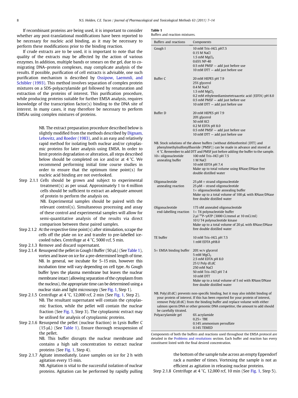
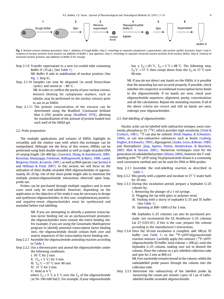
分享
Principles and problems of the electrophoretic mobility shift assay(1).pdf
下载文档

ID:3616328

大小:632.49KB

页数:8页

格式:PDF

时间:2024-06-26

收藏 分享赚钱
温馨提示:
1. 部分包含数学公式或PPT动画的文件,查看预览时可能会显示错乱或异常,文件下载后无此问题,请放心下载。
2. 本文档由用户上传,版权归属用户,汇文网负责整理代发布。如果您对本文档版权有争议请及时联系客服。
3. 下载前请仔细阅读文档内容,确认文档内容符合您的需求后进行下载,若出现内容与标题不符可向本站投诉处理。
4. 下载文档时可能由于网络波动等原因无法下载或下载错误,付费完成后未能成功下载的用户请联系客服处理。
网站客服:3074922707
Principles and problems of the electrophoretic mobility shift assay1 assay
Original articlePrinciples and problems of the electrophoretic mobility shift assayNeil S.Holdena,Claire E.TaconbaDepartment of Cell Biology and Anatomy,Airway Inflammation Research Group,Faculty of Medicine,University of Calgary,Calgary,Alberta,Canada T2N 4N1bDepartment of Physiology and Pharmacology,Airway Inflammation Research Group,Faculty of Medicine,University of Calgary,Calgary,Alberta,Canada T2N 4N1a b s t r a c ta r t i c l ei n f oArticle history:Received 31 December 2009Accepted 11 March 2010Keywords:EMSAMethodsSupershiftDNARadiolabelledEnd-labellingTroubleshooting32PAutoradiographyIntroduction:The electrophoretic mobility shift assay(EMSA)is classically used to detect DNA bindingproteins,the tenet of the EMSA is that DNA with protein bound,migrates through a polyacrylamide gel moreslowly than the corresponding free unbound DNA.Methods:The classical EMSA protocol has 4 major steps:1)The isolation of proteins from cells.Since the vast majority of active DNA binding proteins are present withinthe nucleus,a sequential membrane lysis protocol is used which yields purified nuclear protein.2)Manufactureand radiolabelling of the DNA probe.Phosphorous 32(32P)is attached to the 5 ends of the DNA probe throughuse of32P-ATP as a substrate for T4 polynucleotide kinase.DNA probes can both bepurchasedor custom made.3)Purified proteins and radiolabelled DNA probes are co-incubated with an EMSA binding buffer to promotebinding of the proteinswith the DNA probe.Ifa supershiftEMSA isbeingcarriedout,the reaction also contains aselective antibody which when bound to the proteinDNA complexes,causes further retardation within the gel.4)TheDNAproteincomplexesareloadedandrunonanon-denaturingpolyacrylamidegelcausingseparationofthe DNAprotein complexes from the free DNA probes.The polyacrylamide gels are then dried down andanalysed via autoradiography.Results:As a demonstration of the effectiveness of this protocol,we show thattumournecrosisfactor(TNF)andphorbol12-myristate13-acetate(PMA)stimulationofA549cells,results inanumberof DNAprotein complexes being inducedwhencomparedto untreated cells.We also demonstratethatthese complexes contain the p50 and p65 subunits of NF-B through utilisation of the EMSA supershift protocol.Discussion:We provide detailed troubleshooting hints and tips for this technique and discuss the limitations ofthe EMSA,as well as a number of EMSA variants and alternative techniques.2010 Elsevier Inc.All rights reserved.1.IntroductionThe electrophoretic mobility shift assay(EMSA)was established asa method to detect DNA binding proteins(Fried&Crothers,1981).The principle being that a nucleic acid with protein bound,has lessmobility through a gel matrix than free nucleic acid.One of theprimary applications of the EMSA,is the detection of transcriptionfactors and other sequence specific DNA binding proteins.Althoughnumerous variations on the EMSA have since been developed,each ofthem shares the same basic premise.As one might expect for such awell established technique,there are a number of reviews which havepreviously been published with varying degrees of detail.For anoverview of the EMSA technique we recommend Newton and Staples(2001)and Hellman and Fried(2007).In this current review we haveprovided a detailed step by step guide of the“classical”radioisotopelabelled probe,DNA EMSA.2.Problems and resolutionsIn the following section we present a protocol for the electropho-retic mobility shift assay that we have developed through many yearsof research.At each step,as appropriate,we have included hints,tipsand explanations which may help in the resolution of any problemsthat may arise.We also include a troubleshooting section to detail thesteps which can be taken should the below technique not produce thedesired results.2.1.Preparation of protein samplesTheDNAEMSAcanbe broadlydividedintotwocategories,thoseinwhich the nucleic acid binding proteins identity and properties areknown,and those EMSAs in which the protein binding to nucleic acidsis unknown.Thus the correct preparation of protein samples is vitallyimportant to the successful performance of an EMSA.Crude proteinextracts,partially purified proteins and recombinant or purifiedproteins may be used.The application of the assay will dictate theprotein sample preparation and the optimal protein concentrationneeded.Journal of Pharmacological and Toxicological Methods 63(2011)714 Corresponding author.Department of Cell Biology and Anatomy,Faculty ofMedicine,University of Calgary,3330 Hospital Drive NW,Calgary,Alberta,CanadaT2N 4N1.Tel.:+1 403 220 6329.E-mail address:nholdenucalgary.ca(N.S.Holden).1056-8719/$see front matter 2010 Elsevier Inc.All rights reserved.doi:10.1016/j.vascn.2010.03.002Contents lists available at ScienceDirectJournal of Pharmacological and Toxicological Methodsjournal homepage: recombinant proteins are being used,it is important to considerwhether any post-translational modifications have been reported tobe necessary for nucleic acid binding,as it may be necessary toperform these modifications prior to the binding reaction.If crude extracts are to be used,it is important to note that thequality of the extracts may be affected by the action of variousenzymes.In addition,multiple bands or smears on the gel,due to co-migrating DNAprotein complexes,may complicate analysis of theresults.If possible,purification of cell extracts is advisable,one suchpurification mechanism is described by Ossipow,Laemmli,andSchibler(1993).This method involves separation of complex proteinmixtures on a SDS-polyacrylamide gel followed by renaturation andextraction of the proteins of interest.This purification procedure,while producing proteins suitable for further EMSA analysis,requiresknowledge of the transcription factor(s)binding to the DNA site ofinterest.In many cases,it may therefore be necessary to performEMSAs using complex mixtures of proteins.NB.The extract preparation procedure described below isslightly modified from the methods described by Dignam,Lebovitz,and Roeder(1983),and is an easy and relativelyrapid method for isolating both nuclear and/or cytoplas-mic proteins for later analysis using EMSA.In order tolimit protein degradation or alteration,all steps describedbelow should be completed on ice and/or at 4 C.Werecommend performing initial time course studies inorder to ensure that the optimum time point(s)fornucleic acid binding are not overlooked.Step 2.1.1 Cells should be grown and subject to experimentaltreatment(s)as per usual.Approximately 1 to 4 millioncells should be sufficient to extract an adequate amountof protein to perform the analysis on.NB.Experimental samples should be paired with therelevant control(s).Simultaneous processing and assayof these control and experimental samples will allow forsemi-quantitative analysis of the results via directcomparison between these paired samples.Step 2.1.2 Attherespectivetimepoint(s)afterstimulation,scrapethecells off the plate on ice and transfer to pre-labelled ice-cooled tubes.Centrifuge at 4 C,5000 rcf,5 min.Step 2.1.3 Remove and discard supernatant.Step 2.1.4 ResuspendthepelletinGoughIBuffer(50 L)(SeeTable1),vortex and leave on ice for a pre-determined length of time.NB.In general,we incubate for 515 min,however thisincubation time will vary depending on cell type.As Goughbuffer lyses the plasma membrane but leaves the nuclearmembraneintact(allowingseparationofthecytoplasmfromthenucleus),theappropriatetimecanbedeterminedusinganuclear stain and light microscopy(See Fig.1,Step 1).Step 2.1.5 Centrifuge at 4 C,12,000 rcf,2 min(See Fig.1,Step 2).NB.The resultant supernatant will contain the cytoplas-mic fraction,while the pellet will contain the nuclearfraction(See Fig.1,Step 3).The cytoplasmic extract maybe utilised for analysis of cytoplasmic proteins.Step 2.1.6 Resuspend the pellet(nuclear fraction)in Lysis Buffer C(15 L)(See Table 1).Ensure thorough resuspension ofthe pellet.NB.This buffer disrupts the nuclear membrane andcontains a high salt concentration to extract nuclearproteins(See Fig.1,Step 4).Step 2.1.7 Agitate immediately.Leave samples on ice for 2 h withagitation every 15 min.NB.Agitation is vital to the successful isolation of nuclearproteins.Agitation can be performed by rapidly pullingthebottomofthesampletubeacrossanemptyEppendorfrack a number of times.Vortexing the sample is not asefficient as agitation in releasing nuclear proteins.Step 2.1.8 Centrifuge at 4 C,12,000 rcf,10 min(See Fig.1,Step 5).Table 1Buffers and reaction mixtures.Buffers and reactionsComponentsGough I10 mM TrisHCL pH7.50.15 M NaCl1.5 mM MgCl20.65%NP-400.5 mM PMSF add just before use10 mM DTT add just before useBuffer C20 mM HEPES pH 7.925%glycerol0.4 M NaCl1.5 mM MgCl20.2 mM ethylenediaminetetraacetic acid(EDTA)pH 8.00.5 mM PMSF add just before use10 mM DTT add just before useBuffer D20 mM HEPES pH 7.920%glycerol50 mM KCl0.2 M EDTA pH 8.00.5 mM PMSF add just before use10 mM DTT add just before useNB.Stock solutions of the above buffers(without dithiothreitol(DTT)andphenylmethylsulfonylfluoride(PMSF)can be made in advance and stored at4 C.Remember to add DTT and PMSF just before adding the buffer to the sample.10 oligonucleotideannealing buffer100 mM TrisHCl pH 7.51 M NaCl10 mM EDTA pH 7.4Make up to total volume using RNase/DNase freedouble distilled waterOligonucleotideannealing reaction25 M+strand oligonucleotide25 Mstrand oligonucleotide1 oligonucleotide annealing bufferMake up to a total volume of 100 L with RNase/DNasefree double distilled waterOligonucleotideend-labelling reaction175 nM annealed oligonucleotide1 T4 polynucleotide buffer2 l32P-ATP(3000 Ci/mmol at 10 mCi/ml)10 U T4 polynucleotide kinaseMake up to a total volume of 20 L with RNase/DNasefree double distilled waterTE buffer10 mM TrisHCL pH 7.51 mM EDTA pH8.05 EMSA binding buffer20%w/v glycerol5 mM MgCl22.5 mM EDTA pH 8.025 U Poly dI;dC250 mM NaCl50 mM TrisHCl pH 7.410 mM DTTMake up to a total volume of 5 ml with RNase/DNasefree double distilled waterNB.Poly(dI:dC)prevents non-specific binding,but it may also inhibit binding ofyour protein of interest.If this has been reported for your protein of interest,remove Poly(dI:dC)from the binding buffer and replace volume with eithersalmon sperm DNA or other genomic DNA competitor,the amount to add shouldbe carefully titrated.Polyacrylamide gel6%acrylamide0.25 TBE0.14%ammonium persulfate0.14%TEMEDComponents of both the buffers and reactions used throughout the EMSA protocol aredetailed in the Problems and resolutions section.Each buffer and reaction has everyconstituent listed with the final desired concentration.8N.S.Holden,C.E.Tacon/Journal of Pharmacological and Toxicological Methods 63(2011)714Step 2.1.9 Transfer supernatant to a new ice-cooled tube containingBuffer D(35 L)(See Table 1).NB.Buffer D aids in stabilisation of nuclear proteins(SeeFig.1,Step 6).Step 2.1.10 Samples can now be aliquoted(to avoid freeze/thawcycles)and stored at 80 C.NB.In order to confirm the purity of your nuclear extract,western blotting for cytoplasmic markers,such astubulin,may be performed on the nuclear extracts priorto use in an EMSA.Step 2.1.11 The protein concentration of the extracts can bedetermined using the Bradford(Coomassie brilliantblue G-250)protein assay(Bradford,1976),allowingfor standardisation of the amount of protein loaded intoeach well of the EMSA gel.2.2.Probe preparationThe multiple applications and variants of EMSA,highlight itsversatility and the relative ease with which this technique can bemanipulated.Although not the focus of this review,EMSAs can beperformed using both double-stranded as well as single-stranded DNAof varying length(20400 bp)(Gurevich,Zhang,&Aneskievich,2005;Kironmai,Muniyappa,Friedman,Hollingsworth,&Byers,1998;Laniel,Bergeron,Poirier,&Guerin,1997),as well asRNA species(see Section4and Hellman&Fried,2007).In this section,we will focus on theutilisation of short double-stranded DNA oligonucleotides of approxi-mately 2025 bp.Use of this short-probe length aids to minimise themultiple protein/oligonucleotide interactions observed upon long-probe use.Probes can be purchased through multiple suppliers and in mostcases need only be end-labelled.However,depending on theapplication or the objective of the study it may be necessary to designand synthesise oligonucleotides.In this case,complimentary positive-and negative-sense oligonucleotides must be synthesised andannealed before end-labelling.NB.If you are attempting to identify a putative transcrip-tion factor binding site on an uncharacterised promoter,the oligonucleotides must contain the entire binding site.For example,if you are using the Genomatix MatInspectorprogram to identify potential transcription factor bindingsites,the oligonucleotide should contain both core andmatrix sequences of the transcription factor binding site.Step 2.2.1 Assemble the oligonucleotide annealing reaction accordingto Table 1.Step 2.2.2 Use a thermocycler and anneal the oligonucleotides underthe following conditions:I.95 C for 2 minII.(Tm+5 C)for 5 minIII.TmC37 C over 90 minIV.37 C for 2 minV.Hold at 4 Cwhere Tm+5 C is 5 C over the Tmof the oligonucleotide(at 50100 mM NaCl).For example,if your oligonucleotidehas a Tm=83 C,Tm+5 C=88 C.The following step,TmC37 C then ramps down from the Tmto 37 C over90 min.NB.If you do not detect any bands on the EMSA,it is possiblethat the annealing has not occured properly.If possible,checkwhetherthe respective recombinant transcription factor bindsto the oligonucleotide.If no bands are seen,check youroligonucleotide sequences,alignment,purity,concentrationsand all the calculations.Repeat the annealing reaction.If all ofthe above criteria are correct and still no bands are seen,redesign your oligonucleotides.2.3.End-labelling of oligonucleotidesNucleic acids can be labelled with radioactive isotopes,most com-monly phosphorus 32(32P),which provides high sensitivity(Fried&Crothers,1981).33P can also be utilised(Wolf,Hopley,&Schweizer,1994),as can non-radioactive substances such as biotin(Ludwig,Hughes,&Schwartz,1995),digoxygenin(Suske,Gross,&Beato,1989)and fluorophores(Jing,Agnew,Patton,Hendrickson,&Beechem,2003;Man&Stormo,2001).Numerous techniques exist for thegenerationofradiolabelledprobes(Hilario,2004),howeverthe5end-labelling with32P-ATP using T4 polynucleotide kinase is a commonlyused convenient method and can be used for DNA or RNA probes.Step 2.3.1 Assemble the end-labelling reaction as described inTable 1.Step 2.3.2 Mix gently with a pipette and incubate in 37 C water bathfor 30 min.Step 2.3.3 During this incubation period,prepare a Sephadex G-25column by;I.Removing the plunger of a 1 ml syringe.II.Plugging the tip with glass or polymer wool.III.Packing with a slurry of sephadex G-25 and TE buffer(See Table 1).IV.Spinning at 8001000 rcf for 5 min.NB.Sephadex G-25 columns can also be purchased pre-made(we recommend the GE Healthcare G-25 columnsCat 27-5325-01).If this is the case,prepare the columnaccord

此文档下载收益归作者所有

下载文档
你可能关注的文档
收起
展开