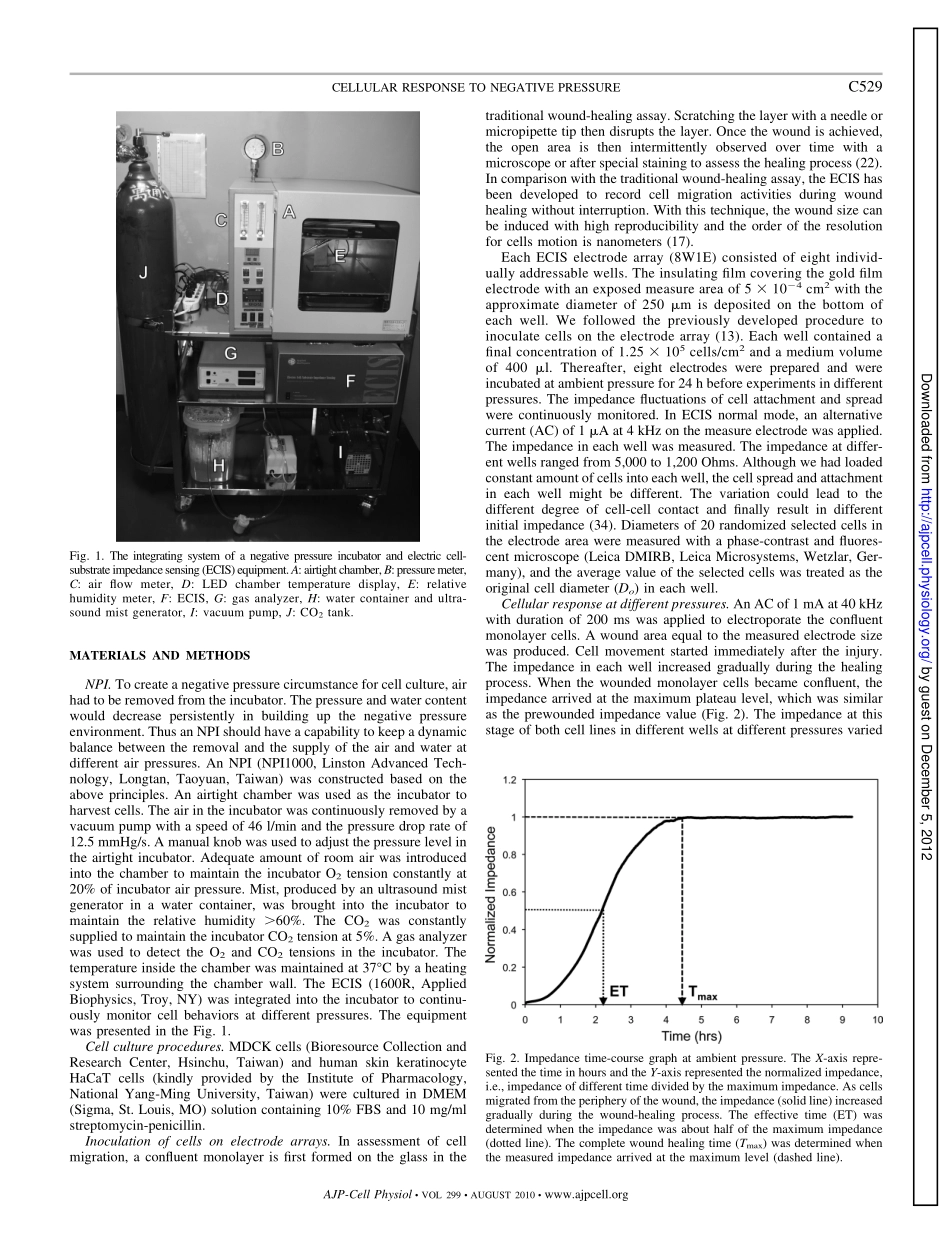
分享
Effects of Negative Pressures on Epithelial Tight Junctions and Migration in Wound Healing(1).pdf
下载文档

ID:3616202

大小:1.68MB

页数:7页

格式:PDF

时间:2024-06-26

收藏 分享赚钱
温馨提示:
1. 部分包含数学公式或PPT动画的文件,查看预览时可能会显示错乱或异常,文件下载后无此问题,请放心下载。
2. 本文档由用户上传,版权归属用户,汇文网负责整理代发布。如果您对本文档版权有争议请及时联系客服。
3. 下载前请仔细阅读文档内容,确认文档内容符合您的需求后进行下载,若出现内容与标题不符可向本站投诉处理。
4. 下载文档时可能由于网络波动等原因无法下载或下载错误,付费完成后未能成功下载的用户请联系客服处理。
网站客服:3074922707
Effects of Negative Pressures on Epithelial Tight Junctions and Migration in Wound Healing1 Healing
Effects of negative pressures on epithelial tight junctions and migration inwound healingChih-Chin Hsu,1,2,3Wen-Chung Tsai,2,3,4Carl Pai-Chu Chen,4Yun-Mei Lu,3and Jong-Shyan Wang31Department of Physical Medicine and Rehabilitation,Chang Gung Memorial Hospital at Keelung,Keelung;2School ofTraditional Chinese Medicine,Chang Gung University,Taoyuan;3Graduate Institute of Rehabilitation Science,College ofMedicine,Chang Gung University,Taoyuan,and4Department of Physical Medicine and Rehabilitation,Chang GungMemorial Hospital at Linkou,Taoyuan,TaiwanSubmitted 12 November 2009;accepted in final form 30 April 2010Hsu CC,Tsai WC,Chen CP,Lu YM,Wang JS.Effects ofnegative pressures on epithelial tight junctions and migration inwound healing.Am J Physiol Cell Physiol 299:C528C534,2010.First published May 5,2010;doi:10.1152/ajpcell.00504.2009.Neg-ative-pressure wound therapy has recently gained popularity inchronic wound care.This study attempted to explore effects ofdifferent negative pressures on epithelial migration in the wound-healing process.The electric cell-substrate impedance sensing(ECIS)technique was used to create a 5?10?4cm2wound in the Madin-Darby canine kidney(MDCK)and human keratinocyte(HaCaT)cells.The wounded cells were cultured in a negative pressure incubator atambient pressure(AP)and negative pressures of 75 mmHg(NP75),125 mmHg(NP125),and 175 mmHg(NP175).The effective time(ET),complete wound healing time(Tmax),healing rate(Rheal),cell diam-eter,and wound area over time at different pressures were evaluated.Traditional wound-healing assays were prepared for fluorescent stain-ing of cells viability,cell junction proteins,including ZO-1 andE-cadherin,and actins.Amount of cell junction proteins at AP andNP125was also quantified.In MDCK cells,the ET(1.25?0.27 h),Tmax(1.76?0.32 h),and Rheal(2.94?0.62?10?4cm2/h)at NP125were significantly(P?0.01)different from those at three otherpressure conditions.In HaCaT cells,the Tmax(7.34?0.29 h)andRheal(6.82?0.26?10?5cm2/h)at NP125were significantly(P?0.01)different from those at NP75.Prominent cell migration featureswere identified in cells at the specific negative pressure.Cell migra-tion activities at different pressures can be documented with thereal-time wound-healing measurement system.Negative pressure of125 mmHg can help disassemble the cell junction to enhance epithe-lial migration and subsequently result in quick wound closure.negative-pressure wound therapy;electric impedance;cell junctions;cell movementAN ESTIMATED THREE MILLIONindividuals are believed to havechronic wounds and billions of dollars are spent on treatmenteach year in the United States(12).The lifetime risk ofdeveloping foot ulcers in the diabetic patient may be as high as25%(26).Venous ulcers or wounds resulting from arterialdisease may also lead to chronic wound problems(14).Dif-ferent types of dressings are developed but few of them haveconvincingly been shown to provide higher wound closurerates than traditional wet gauze dressings(31).Despite theadvance of current treatments for chronic wounds,many ofthem fail to heal and persist for months to years(25).Toaccelerate wound healing,the negative-pressure wound ther-apy(NPWT)has been developed(20)and has become widelyused in chronic wound cares(30).A significant reduction of the partial foot amputation woundsize in patients treated with NPWT has been reported in arandomized control trial(1).Another trial has demonstratedthat this kind of therapy could increase granulation tissueformation in chronic nonhealing wounds(12).The negativepressure of 125 mmHg has been used as a therapeutic dosagein clinical practice based on the subcutaneous flow measure-ments in a swine study(20).However,investigators haveconcluded that negative pressure of 125 mmHg might not bethe optimal pressure for wound healing based on the cutaneousblood flow measurement in 10 healthy subjects(29).A nega-tive pressure of 75 mmHg has also been found to work onholding a skin graft in place(11).Thus exploring mechanismsfor effects of the negative pressure on wound healing isrequired to clarify the controversial findings.With the aid of a commercialized NPWT device,differentnegative pressures have been applied on human endothelial cells(2)and human dermal fibroblasts(19).Conflicted results regard-ing cell viability and migration response to negative pressure havebeen observed in the previous reports.These laboratory findingsdid not provide strong support for the encouraging clinical obser-vations of NPWT for chronic wounds(1,12).Wound-healing assays have been carried out in cell cultures formany years to monitor the healing process.A new method ofobtaining cell migration information using an electrical means toassess the wound-healing process is referred as electric cell-substrate impedance sensing(ECIS).As cells grow and move onthe ECIS measure electrode,the impedance fluctuates until thecell layer becomes confluent.The value is directly proportional tothe cell coverage(9)and intercellular resistance(34).The ECIStechnique has been used to continuously monitor Madin-Darbycanine kidney(MDCK)epithelial cell attachment and spreading(34),the relationship between calcium signaling and coherentchanges in adhesion properties of hormone-stimulated MDCKcells(7),and effects of exercise on natural killer cell activities(33).Previous studies have also used the technique to evaluate theimpedance change along time of human keratinocytes to differentchemical toxicants(6),the adhesion capacity of normal humankeratinocytes to different adhesive peptides(21),and the keratin-ocyte migration response to the epidermal growth factor(37).Therefore,the ECIS can be an ideal choice for in-depth analysisof cell behaviors under various conditions.The purpose of thepresent work is to integrate a negative pressure incubator(NPI)and the ECIS technique to continuously observe the healingprocess of wounded monolayer cells at different negative pres-sures.Address for reprint requests and other correspondence:J.-S.Wang,GraduateInstitute of Rehabilitation Science,Chang Gung Univ.,259,Wen-Hwa 1stRoad,Kwei-Shan,Taoyuan 333,Taiwan(e-mail:s5492mail.cgu.edu.tw).Am J Physiol Cell Physiol 299:C528C534,2010.First published May 5,2010;doi:10.1152/ajpcell.00504.2009.0363-6143/10 Copyright2010 the American Physiological Societyhttp:/www.ajpcell.orgC528 by guest on December 5,2012http:/ajpcell.physiology.org/Downloaded from MATERIALS AND METHODSNPI.To create a negative pressure circumstance for cell culture,airhad to be removed from the incubator.The pressure and water contentwould decrease persistently in building up the negative pressureenvironment.Thus an NPI should have a capability to keep a dynamicbalance between the removal and the supply of the air and water atdifferent air pressures.An NPI(NPI1000,Linston Advanced Tech-nology,Longtan,Taoyuan,Taiwan)was constructed based on theabove principles.An airtight chamber was used as the incubator toharvest cells.The air in the incubator was continuously removed by avacuum pump with a speed of 46 l/min and the pressure drop rate of12.5 mmHg/s.A manual knob was used to adjust the pressure level inthe airtight incubator.Adequate amount of room air was introducedinto the chamber to maintain the incubator O2tension constantly at20%of incubator air pressure.Mist,produced by an ultrasound mistgenerator in a water container,was brought into the incubator tomaintain the relative humidity?60%.The CO2was constantlysupplied to maintain the incubator CO2tension at 5%.A gas analyzerwas used to detect the O2and CO2tensions in the incubator.Thetemperature inside the chamber was maintained at 37C by a heatingsystem surrounding the chamber wall.The ECIS(1600R,AppliedBiophysics,Troy,NY)was integrated into the incubator to continu-ously monitor cell behaviors at different pressures.The equipmentwas presented in the Fig.1.Cell culture procedures.MDCK cells(Bioresource Collection andResearch Center,Hsinchu,Taiwan)and human skin keratinocyteHaCaT cells(kindly provided by the Institute of Pharmacology,National Yang-Ming University,Taiwan)were cultured in DMEM(Sigma,St.Louis,MO)solution containing 10%FBS and 10 mg/mlstreptomycin-penicillin.Inoculation of cells on electrode arrays.In assessment of cellmigration,a confluent monolayer is first formed on the glass in thetraditional wound-healing assay.Scratching the layer with a needle ormicropipette tip then disrupts the layer.Once the wound is achieved,the open area is then intermittently observed over time with amicroscope or after special staining to assess the healing process(22).In comparison with the traditional wound-healing assay,the ECIS hasbeen developed to record cell migration activities during woundhealing without interruption.With this technique,the wound size canbe induced with high reproducibility and the order of the resolutionfor cells motion is nanometers(17).Each ECIS electrode array(8W1E)consisted of eight individ-ually addressable wells.The insulating film covering the gold filmelectrode with an exposed measure area of 5?10?4cm2with theapproximate diameter of 250?m is deposited on the bottom ofeach well.We followed the previously developed procedure toinoculate cells on the electrode array(13).Each well contained afinal concentration of 1.25?105cells/cm2and a medium volumeof 400?l.Thereafter,eight electrodes were prepared and wereincubated at ambient pressure for 24 h before experiments in differentpressures.The impedance fluctuations of cell attachment and spreadwere continuously monitored.In ECIS normal mode,an alternativecurrent(AC)of 1?A at 4 kHz on the measure electrode was applied.The impedance in each well was measured.The impedance at differ-ent wells ranged from 5,000 to 1,200 Ohms.Although we had loadedconstant amount of cells into each well,the cell spread and attachmentin each well might be different.The variation could lead to thedifferent degree of cell-cell contact and finally result in differentinitial impedance(34).Diameters of 20 randomized selected cells inthe electrode area were measured with a phase-contrast and fluores-cent microscope(Leica DMIRB,Leica Microsystems,Wetzlar,Ger-many),and the average value of the selected cells was treated as theoriginal cell diameter(Do)in each well.Cellular response at different pressures.An AC of 1 mA at 40 kHzwith duration of 200 ms was applied to electroporate the confluentmonolayer cells.A wound area equal to the measured electrode sizewas produced.Cell movement started immediately after the injury.The impedance in each well increased gradually during the healingprocess.When the wounded monolayer cells became confluent,theimpedance arrived at the maximum plateau level,which was similaras the prewounded impedance value(Fig.2).The impedance at thisstage of both cell lines in different wells at different pressures variedFig.1.The integrating system of a negative pressure incubator and electric cell-substrateimpedancesensing(ECIS)equipment.A:airtightchamber,B:pressuremeter,C:air flow meter,D:LED chamber temperature display,E:relativehumidity meter,F:ECIS,G:gas analyzer,H:water container and ultra-sound mist generator,I:vacuum pump,J:CO2tank.Fig.2.Impedance time-course graph at ambient pressure.The X-axis repre-sented the time in hours and the Y-axis represented the normalized impedance,i.e.,impedance of different time divided by the maximum impedance.As cellsmigrated from the periphery of the wound,the impedance(solid line)increasedgradually during the wound-healing process.The effective time(ET)wasdetermined when the impedance was about half of the maximum impedance(dotted line).The complete wound healing time(Tmax)was determined whenthe measured impedance arrived at the maximum level(dashed line).C529CELLULAR RESPONSE TO NEGATIVE PRESSUREAJP-Cell PhysiolVOL 299AUGUST 2010www.ajpcell.org by guest on December 5,2012http:/ajpcell.physiology.org/Downloaded from from 5,500 to 15,000 Ohms.To compare resistance between differentwells at different pressures,the relative degree of impedance,i.e.,impedance at certain point during the wound-healing process wasdivided by the maximum impedance at complete wound healing stage,was used in the study.The effective time(ET),representing thesensitivity of wounded cells to the applied pressure,was defined as thetime between the immediately postwounded point and the time at 50%of the maximum impedance.The time from the beginning of woundhealing to the time of the maximum impedance(Tmax)was defined ascomplete wound healing time.The shorter the two parameters werethe faster the wound healed.The wounded cells in 16 wells in twocontrol electrode arrays were cultured in our self-constructed incuba-tor at ambient pressure(AP).Every two of the other six electrodearrays were harvested under the negative pressure of 75(NP75),125(NP125),and 175(NP175)mmHg,respectively.Therefore,the testedsample number was 16 in each group.Healing processes at differentpressures were shown in the Fig.3.The monolayer cell wound healingrate(Rheal)was estimated as 5?10?4/Tmaxcm2/h.Each well at ETand Tmaxwas examined with the microscope.The cell diameter ineach well at the complete wound-healing stage(Dend)at differentpressures was determined as the measurement for Do.The strain()ofcells at different pressures could be derived as(Dend?D0)/D0.Thewound area(A)in each well at ET in the four groups was measuredwith Photoshop CS4 extended(Adobe System,San Jose,CA).The Awas further normalized with the measure electrode area(AN)and wascalculated as A/5?10?4.Therefore,the ANat initial and endwound-healing process would always be 1 and 0,respectively.TheET,Tmax,Rheal,Do,Dend,and ANin the four groups were recorded.Traditional wound healing assay.A 400-?l DMEM medium con-taining 1.25?105cells/cm2was inoculated on each well in thechambered coverglass system(Lab-Tek,Nalge Nunc International,Naperville,IL),and then the system was incubated overnight.Everycoverglass has eight wells.A pipette tip(0.110?l,Labcon NorthAmerica,Petaluma,CA)was later used to create a scratch wound ineach well.Six prepared coverglasses were then harvested at AP andNP125,respectively,for 3 h.Two of them were prepared for cells vitalstain.The remaining four coverglasses were fixed with 4%parafor-maldehyde for 5 min and prepared for tight junction and actinfilaments stains at room temperatures.There were eight wells in eachstain,respectively,at AP and NP125.Vital staining.The Live/Deads Viability/Cytotoxicity kit(Invitro-gen,Carlsbad,CA)was used for vital staining of cells in twocoverglasses at the two different pressures.The kit differentially stainslive and dead cells.Green fluorescence is indicative of live cells,andred fluorescence indicates dead cells(23).Random selections of fourfields of view at?200,about 8,000 cells in each view,were countedper well with MetaMorph 5.0 software(Molecular Devices,Sunny-vale,CA).The dead cell percentage was determined as numbers ofdead cells divided by total cells in each view.Cell ju

此文档下载收益归作者所有

下载文档
你可能关注的文档
收起
展开