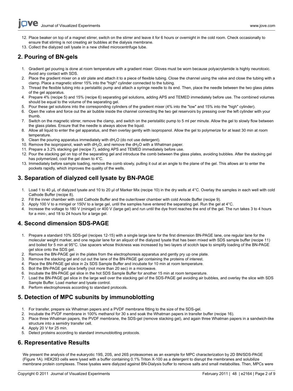
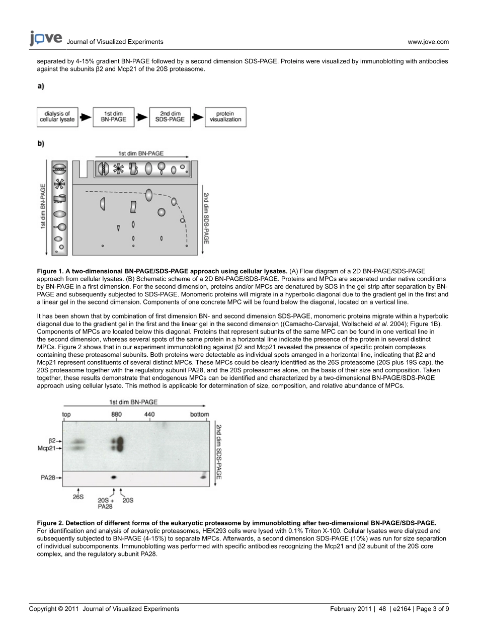
分享
protocol.pdf
下载文档
下载文档

ID:3616097

大小:176.71KB

页数:9页

格式:PDF

时间:2024-06-26

收藏 分享赚钱
温馨提示:
1. 部分包含数学公式或PPT动画的文件,查看预览时可能会显示错乱或异常,文件下载后无此问题,请放心下载。
2. 本文档由用户上传,版权归属用户,汇文网负责整理代发布。如果您对本文档版权有争议请及时联系客服。
3. 下载前请仔细阅读文档内容,确认文档内容符合您的需求后进行下载,若出现内容与标题不符可向本站投诉处理。
4. 下载文档时可能由于网络波动等原因无法下载或下载错误,付费完成后未能成功下载的用户请联系客服处理。
网站客服:3074922707
protocol
Journal of Visualized ECopyright 2011 Journal of Visualized ExperimentsFebruary 2011|48|e2164|Page 1 of 9Video ArticleBlue Native Polyacrylamide Gel Electrophoresis(BN-PAGE)for Analysis ofMultiprotein Complexes from Cellular LysatesGina J.Fiala1,2,3,Wolfgang W.A.Schamel*2,3,Britta Blumenthal*31Spemann Graduate School of Biology and Medicine(SGBM),University of Freiburg2Centre for Biological Signalling Studies(bioss)and Biology III,Faculty of Biology,University of Freiburg3Department of Molecular Immunology,Max-Planck-Institute of Immunology and Epigenetics*These authors contributed equallyCorrespondence to:Wolfgang W.A.Schamel at schamelimmunbio.mpg.de,Britta Blumenthal at blumenthalimmunbio.mpg.deURL:http:/ 48,BN-PAGE,2D gel electrophoresis,cellular lysate,dialysis,protein complex,multiprotein complex,proteininteractionDate Published:2/24/2011Citation:Fiala,G.J.,Schamel,W.W.A.,Blumenthal,B.Blue Native Polyacrylamide Gel Electrophoresis(BN-PAGE)for Analysis of MultiproteinComplexes from Cellular Lysates.J.Vis.Exp.(48),e2164,doi:10.3791/2164(2011).AbstractMultiprotein complexes(MPCs)play a crucial role in cell signalling,since most proteins can be found in functional or regulatory complexeswith other proteins(Sali,Glaeser et al.2003).Thus,the study of protein-protein interaction networks requires the detailed characterization ofMPCs to gain an integrative understanding of protein function and regulation.For identification and analysis,MPCs must be separated undernative conditions.In this video,we describe the analysis of MPCs by blue native polyacrylamide gel electrophoresis(BN-PAGE).BN-PAGE isa technique that allows separation of MPCs in a native conformation with a higher resolution than offered by gel filtration or sucrose densityultracentrifugation,and is therefore useful to determine MPC size,composition,and relative abundance(Schgger and von Jagow 1991);(Schgger,Cramer et al.1994).By this method,proteins are separated according to their hydrodynamic size and shape in a polyacrylamidematrix.Here,we demonstrate the analysis of MPCs of total cellular lysates,pointing out that lysate dialysis is the crucial step to make BN-PAGE applicable to these biological samples.Using a combination of first dimension BN-and second dimension SDS-PAGE,we show thatMPCs separated by BN-PAGE can be further subdivided into their individual constituents by SDS-PAGE.Visualization of the MPC componentsupon gel separation is performed by standard immunoblotting.As an example for MPC analysis by BN-PAGE,we chose the well-characterizedeukaryotic 19S,20S,and 26S proteasomes.Video LinkThe video component of this article can be found at http:/ video protocol is based on an associated publication 1:Blue Native Polyacrylamide Gel Electrophoresis(BN-PAGE)for theIdentification and Analysis of Multiprotein Complexes.Mahima Swamy,Gabrielle M.Siegers,Susana Minguet,Bernd Wollscheid,and WolfgangW.A.Schamel.Sciences STKE 2006(345):pl2,July 25,2006,DOI:10.1126/stke.3892006pl4.Please click here to see this publication.1.Preparation of dialyzed cell lysate1.Harvest 10 x106 cells and pellet by centrifugation at 350g for 5 min at 4C.2.Wash the cell pellet three times with 1 mL of ice-cold PBS(recipe 1),centrifuge as in step 1.1.3.Resuspend pellet in 250 L of ice-cold BN-Lysis Buffer(recipe 2)and incubate on ice for 15 min.4.Centrifuge at 13,000g for 15 min at 4C to remove insoluble material.5.Melt a hole in the cap of a 1.5-mL microcentrifuge tube using the heated large diameter side of a Pasteur pipette,then place the tube on iceto cool down to 4C.6.Transfer supernatant from step 1.4 into the chilled tube with the hole in the cap.7.Place a dialysis membrane(molecular weight cut-off of 10 kD)with forceps on top of the opened tube,close the cap,and cut off excessdialysis membrane that sticks out.8.Seal the cap on the side carefully with Parafilm.9.Invert the tubes and centrifuge upside-down at the lowest speed possible in a 50-mL conical tubes in a cell culture centrifuge for 10 sec at4C.Remove the inverted tube from the centrifuge using tweezers to avoid turning the tube right side up.10.Prepare a 100-mL beaker with cold BN-Dialysis Buffer(recipe 3)and a stir plate.Use at least 10 mL of BN-Dialysis Buffer per 100-L sample.11.Affix the tube with tape upside-down inside the beaker,and remove air bubbles from the hole beneath the cap using a bent Pasteur pipette.Journal of Visualized ECopyright 2011 Journal of Visualized ExperimentsFebruary 2011|48|e2164|Page 2 of 912.Place beaker on top of a magnet stirrer,switch on the stirrer and leave it for 6 hours or overnight in the cold room.Check occasionally toensure that stirring is not creating air bubbles at the dialysis membrane.13.Collect the dialyzed cell lysate in a new chilled microcentrifuge tube.2.Pouring of BN-gels1.Gradient gel pouring is done at room temperature with a gradient mixer.Gloves must be worn because polyacrylamide is highly neurotoxic.Avoid any contact with SDS.2.Place the gradient mixer on a stir plate and attach it to a piece of flexible tubing.Close the channel using the valve and close the tubing with aclamp.Place a magnetic stirrer 15%into the high cylinder connected to the tubing.3.Thread the flexible tubing into a peristalitic pump and attach a syringe needle to its end.Then,place the needle between the two glass platesof the gel apparatus.4.Prepare 4%(recipe 5)and 15%(recipe 6)separating gel solutions,adding APS and TEMED immediately before use.The combined volumesshould be equal to the volume of the separating gel.5.Pour these gel solutions into the corresponding cylinders of the gradient mixer(4%into the low and 15%into the high cylinder).6.Open the valve and force out the air bubble inside the channel connecting the two gel reservoirs by pressing over the left cylinder with yourthumb.7.Switch on the magnetic stirrer,remove the clamp,and switch on the peristalitic pump to 5 ml per minute.Allow the gel to slowly flow betweenthe glass plates.Ensure that the needle is always above the liquid.8.Allow all liquid to enter the gel apparatus,and then overlay gently with isopropanol.Allow the gel to polymerize for at least 30 min at roomtemperature.9.Clean the pouring apparatus immediately with dH2O(do not use detergent).10.Remove the isopropanol,wash with dH2O,and remove the dH2O with a Whatman paper.11.Prepare a 3.2%stacking gel(recipe 7),adding APS and TEMED immediately before use.12.Pour the stacking gel on top of the separating gel and introduce the comb between the glass plates,avoiding bubbles.After the stacking gelhas polymerized,cool the gel down to 4C.13.Immediately before sample loading,remove the comb slowly,pulling it out at an angle to the plane of the gel.This allows air to enter thepockets rapidly,which improves the quality of the wells.3.Separation of dialyzed cell lysate by BN-PAGE1.Load 1 to 40 L of dialyzed lysate and 10 to 20 l of Marker Mix(recipe 10)in the dry wells at 4C.Overlay the samples in each well with coldCathode Buffer(recipe 8).2.Fill the inner chamber with cold Cathode Buffer and the outer/lower chamber with cold Anode Buffer(recipe 9).3.Apply 100 V to a minigel or 150V to a large gel,until the samples have entered the separating gel.Run the gel at 4C.4.Increase the voltage to 180 V(minigel)or 400 V(large gel)and run until the dye front reaches the end of the gel.The run takes 3 to 4 hoursfor a mini-,and 18 to 24 hours for a large gel.4.Second dimension SDS-PAGE1.Prepare a standard 10%SDS-gel(recipes 12-15)with a single large lane for the first dimension BN-PAGE lane,one regular lane for themolecular weight marker,and one regular lane for an aliquot of the dialyzed lysate that has been mixed with SDS sample buffer(recipe 11)and boiled for 5 min at 95C.Use spacers whose thickness was increased by two layers of scotch tape to simplify loading of the BN-PAGEgel slice onto the SDS gel.2.Remove the BN-PAGE gel in the plates from the electrophoresis apparatus and gently pry up one plate.3.Remove the stacking gel and cut out the lane of the BN-PAGE gel containing the proteins of interest.4.Place the BN-PAGE gel slice in 2x SDS Sample Buffer and incubate for 10 min at room temperature.5.Boil the BN-PAGE gel slice briefly(not more than 20 sec)in a microwave.6.Incubate the BN-PAGE gel slice in the hot SDS Sample Buffer for another 15 min at room temperature.7.Load the BN-PAGE gel slice in the large well over the stacking gel of the SDS-PAGE gel avoiding air bubbles,and overlay the slice with SDSSample Buffer.Load marker and lysate control.8.Perform electrophoresis according to standard protocols.5.Detection of MPC subunits by immunoblotting1.For transfer,prepare six Whatman papers and a PVDF membrane fitting to the size of the SDS-gel.2.Incubate the PVDF membrane in 100%methanol for 30 s and soak the Whatman papers in transfer buffer(recipe 16).3.Place three Whatman papers,the PVDF membrane,the SDS-gel(remove stacking gel),and again three Whatman papers in a sandwich-likestructure into a semidry transfer cell.4.Apply 20 V for 25 min.5.Detect proteins according to standard immunoblotting protocols.6.Representative ResultsWe present the analysis of the eukaryotic 19S,20S,and 26S proteasomes as an example for MPC characterization by 2D BN/SDS-PAGE(Figure 1A).HEK293 cells were lysed with a buffer containing 0.1%Triton X-100 as a detergent to disrupt the membranes and solubilizemembrane protein complexes.These lysates were dialyzed against BN-Dialysis buffer to remove salts and small metabolites.Then,MPCs wereJournal of Visualized ECopyright 2011 Journal of Visualized ExperimentsFebruary 2011|48|e2164|Page 3 of 9separated by 4-15%gradient BN-PAGE followed by a second dimension SDS-PAGE.Proteins were visualized by immunoblotting with antibodiesagainst the subunits 2 and Mcp21 of the 20S proteasome.Figure 1.A two-dimensional BN-PAGE/SDS-PAGE approach using cellular lysates.(A)Flow diagram of a 2D BN-PAGE/SDS-PAGEapproach from cellular lysates.(B)Schematic scheme of a 2D BN-PAGE/SDS-PAGE.Proteins and MPCs are separated under native conditionsby BN-PAGE in a first dimension.For the second dimension,proteins and/or MPCs are denatured by SDS in the gel strip after separation by BN-PAGE and subsequently subjected to SDS-PAGE.Monomeric proteins will migrate in a hyperbolic diagonal due to the gradient gel in the first anda linear gel in the second dimension.Components of one concrete MPC will be found below the diagonal,located on a vertical line.It has been shown that by combination of first dimension BN-and second dimension SDS-PAGE,monomeric proteins migrate within a hyperbolicdiagonal due to the gradient gel in the first and the linear gel in the second dimension(Camacho-Carvajal,Wollscheid et al.2004);Figure 1B).Components of MPCs are located below this diagonal.Proteins that represent subunits of the same MPC can be found in one vertical line inthe second dimension,whereas several spots of the same protein in a horizontal line indicate the presence of the protein in several distinctMPCs.Figure 2 shows that in our experiment immunoblotting against 2 and Mcp21 revealed the presence of specific protein complexescontaining these proteasomal subunits.Both proteins were detectable as individual spots arranged in a horizontal line,indicating that 2 andMcp21 represent constituents of several distinct MPCs.These MPCs could be clearly identified as the 26S proteasome(20S plus 19S cap),the20S proteasome together with the regulatory subunit PA28,and the 20S proteasomes alone,on the basis of their size and composition.Takentogether,these results demonstrate that endogenous MPCs can be identified and characterized by a two-dimensional BN-PAGE/SDS-PAGEapproach using cellular lysate.This method is applicable for determination of size,composition,and relative abundance of MPCs.Figure 2.Detection of different forms of the eukaryotic proteasome by immunoblotting after two-dimensional BN-PAGE/SDS-PAGE.For identification and analysis of eukaryotic proteasomes,HEK293 cells were lysed with 0.1%Triton X-100.Cellular lysates were dialyzed andsubsequently subjected to BN-PAGE(4-15%)to separate MPCs.Afterwards,a second dimension SDS-PAGE(10%)was run for size separationof individual subcomponents.Immunoblotting was performed with specific antibodies recognizing the Mcp21 and 2 subunit of the 20S corecomplex,and the regulatory subunit PA28.Journal of Visualized ECopyright 2011 Journal of Visualized ExperimentsFebruary 2011|48|e2164|Page 4 of 9I.Table of specific reagents(alphabetical order):ReagentCompanyComments6-aminohexanoic acid(-aminocaproic acid)Sigma-Aldrich,Taufkirchen,GermanyThis chemical is an irritant and should behandled with gloves.Acrylamide-bisacrylamide solution(40%),Mix32:1Applichem,Darmstadt,GermanyThis solution is neurotoxic and should behandled with gloves.Bis-trisRoth,Karlsruhe,Germa-ny Brij 96Sigma-Aldrich,Taufkirchen,Germany Coomassie blue G250Serva,Heidelberg,Ger-manyDo not substitute other types of Coomassie dyesuch as Coomassie blue R250 or colloidal Coo-massie blues.DigitoninSigma-Aldrich,Taufkirchen,GermanyDigitonin is toxic.Gloves should be worn whenhandling buffers or samples containing thisdeter-gent.DodecylmaltosideApplichem,Darmstadt,Germany Triton X-100Roth,Karlsruhe,Germa-nyTriton X-100 is toxic.Gloves should be wornwhen handling buffers or samples containingthis detergent.II.Table of specific material and equipment:EquipmentCompanyDialysis membranes(molecular weight cut-off 10 to 50 kD)Roth,Karlsruhe,GermanyGel electrophoresis systemFor example from Bio-Rad,Munich,GermanyGradient mixerSelf-made or commercially available from Bio-Rad,Munich,GermanyPeristaltic pumpAmersham Pharmacia Biotech,Freiburg,GermanyPolyvinylidene difluoride(PVDF)membraneImmobilon-P,Millipore,Eschborn,GermanySemi-dry transfer equipmentFor example from Bio-Rad,Munich,GermanySilicon tubing(3 to 5 mm diameter,1 m length)NeoLab,Heidelberg,GermanyIII.Table of recipes:No.Buffers and solutionsContentComments1Phosphate-Buffered Saline(PBS)Na2HPO4 8.1 mMKH2 PO4 1.5 mM NaCl 138 mM KCl 2.7 mMSolution should be pH 7.4 if pre-pared properly.2BN-Lysis BufferBase buffer Bis-tris 20 mM-aminocaproic acid 500 mM NaCl 20 mM EDTA,pH 8.0 2 mM Glycerol 10%Adjust pH to 7.0 with HCl.Store at4C.Detergent The appropriate detergent must bedetermined empirically and shouldbe the same as that used in theother lysis buffer recipes.Digitonin must be added justbefore use from a 2%stocksolution in dH2O(store in 5-mlaliquots at-20C).Protease and phophatase inhibit-ors should be added immediatelybefore use.Journal of Visualized ECopyright 2011 Journal of Visualized ExperimentsFebruary 2011|48|e2164|Page 5 of 9Digitonin 0.5 to 1.0%or Brij 96 0.1 to 0.5%or Triton X-100 0.1 to 0.5%or Dodecylmaltoside 0.1 to 0.5%Protease and phosphataseinhibitors Aprotinin 10 g/mL Leupeptin 10 g/mL PMSF 1 mM Sodium fluoride 0.5 mM Sodium orthovanadate 0.5 mMUpon addition of sodium orthova-nadate,the buffer will becomeyellowish in color.3BN-Dialy

此文档下载收益归作者所有

下载文档
你可能关注的文档
收起
展开