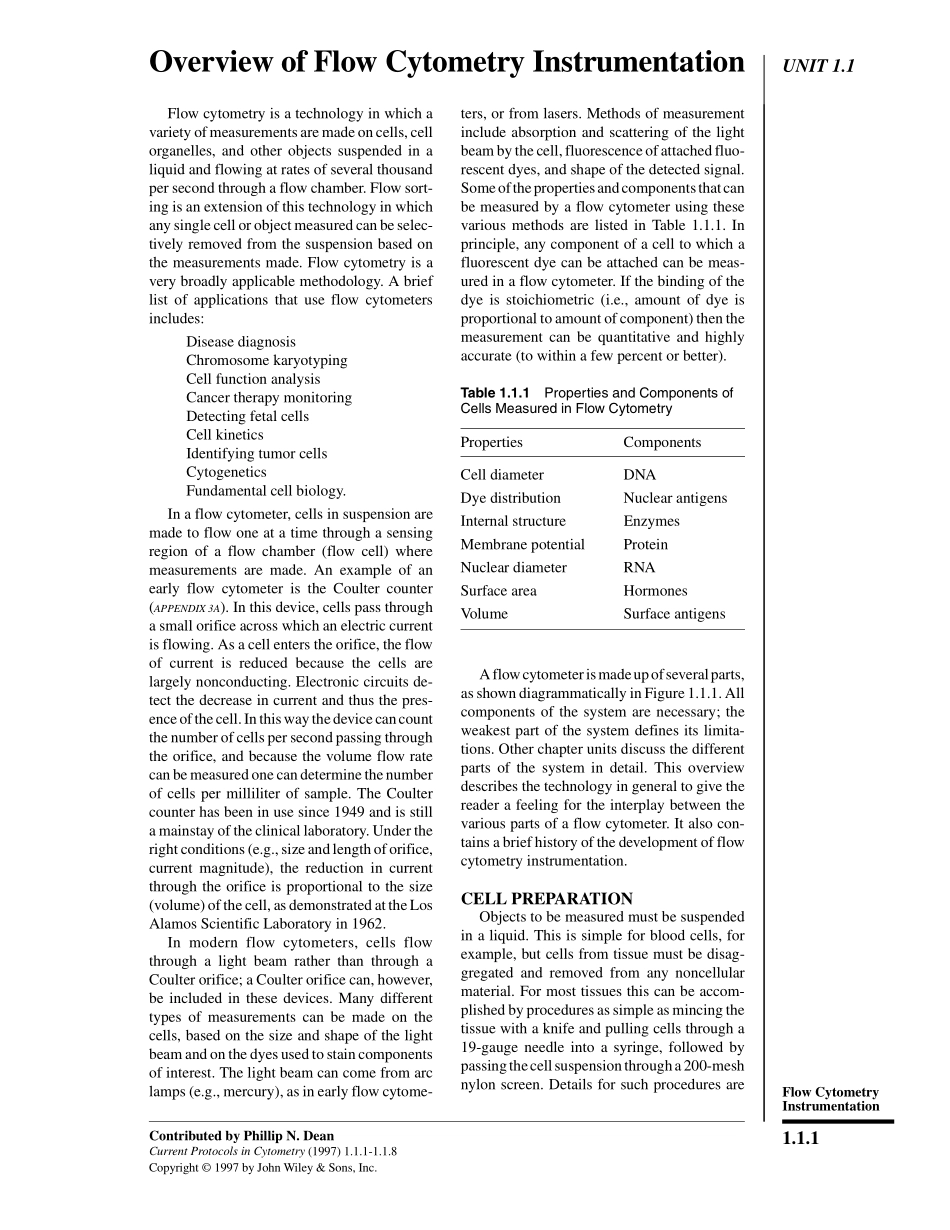
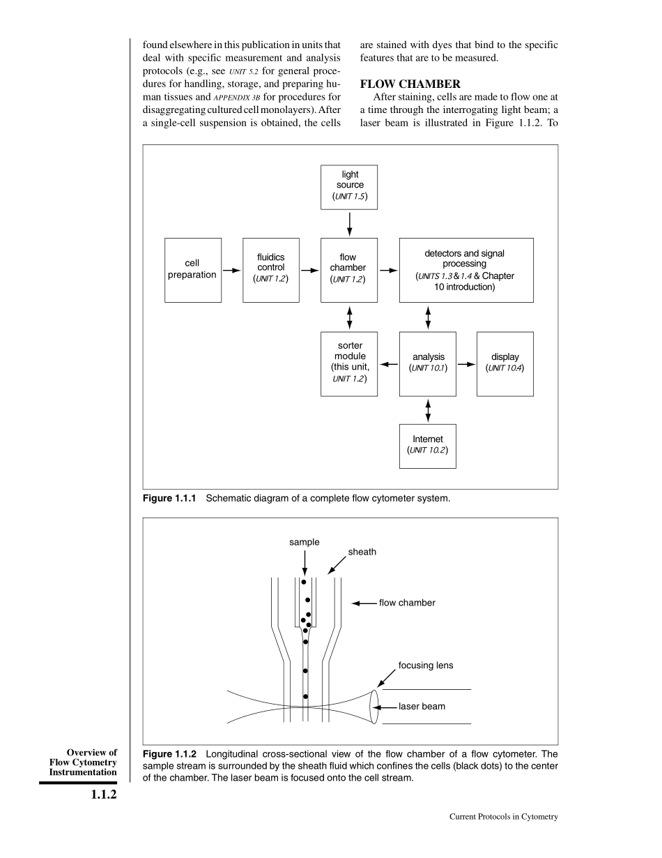
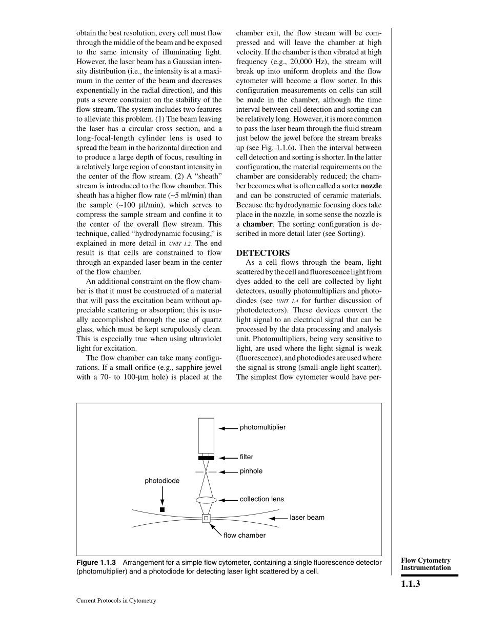
分享
流式细胞术说明.pdf
下载文档

ID:3495862

大小:25.32MB

页数:1981页

格式:PDF

时间:2024-05-16

收藏 分享赚钱
温馨提示:
1. 部分包含数学公式或PPT动画的文件,查看预览时可能会显示错乱或异常,文件下载后无此问题,请放心下载。
2. 本文档由用户上传,版权归属用户,汇文网负责整理代发布。如果您对本文档版权有争议请及时联系客服。
3. 下载前请仔细阅读文档内容,确认文档内容符合您的需求后进行下载,若出现内容与标题不符可向本站投诉处理。
4. 下载文档时可能由于网络波动等原因无法下载或下载错误,付费完成后未能成功下载的用户请联系客服处理。
网站客服:3074922707
细胞 说明
UNIT 1.1Overview of Flow Cytometry InstrumentationFlow cytometry is a technology in which avariety of measurements are made on cells,cellorganelles,and other objects suspended in aliquid and flowing at rates of several thousandper second through a flow chamber.Flow sort-ing is an extension of this technology in whichany single cell or object measured can be selec-tively removed from the suspension based onthe measurements made.Flow cytometry is avery broadly applicable methodology.A brieflist of applications that use flow cytometersincludes:Disease diagnosisChromosome karyotypingCell function analysisCancer therapy monitoringDetecting fetal cellsCell kineticsIdentifying tumor cells CytogeneticsFundamental cell biology.In a flow cytometer,cells in suspension aremade to flow one at a time through a sensingregion of a flow chamber(flow cell)wheremeasurements are made.An example of anearly flow cytometer is the Coulter counter(APPENDIX 3A).In this device,cells pass througha small orifice across which an electric currentis flowing.As a cell enters the orifice,the flowof current is reduced because the cells arelargely nonconducting.Electronic circuits de-tect the decrease in current and thus the pres-ence of the cell.In this way the device can countthe number of cells per second passing throughthe orifice,and because the volume flow ratecan be measured one can determine the numberof cells per milliliter of sample.The Coultercounter has been in use since 1949 and is stilla mainstay of the clinical laboratory.Under theright conditions(e.g.,size and length of orifice,current magnitude),the reduction in currentthrough the orifice is proportional to the size(volume)of the cell,as demonstrated at the LosAlamos Scientific Laboratory in 1962.In modern flow cytometers,cells flowthrough a light beam rather than through aCoulter orifice;a Coulter orifice can,however,be included in these devices.Many differenttypes of measurements can be made on thecells,based on the size and shape of the lightbeam and on the dyes used to stain componentsof interest.The light beam can come from arclamps(e.g.,mercury),as in early flow cytome-ters,or from lasers.Methods of measurementinclude absorption and scattering of the lightbeam by the cell,fluorescence of attached fluo-rescent dyes,and shape of the detected signal.Some of the properties and components that canbe measured by a flow cytometer using thesevarious methods are listed in Table 1.1.1.Inprinciple,any component of a cell to which afluorescent dye can be attached can be meas-ured in a flow cytometer.If the binding of thedye is stoichiometric(i.e.,amount of dye isproportional to amount of component)then themeasurement can be quantitative and highlyaccurate(to within a few percent or better).A flow cytometer is made up of several parts,as shown diagrammatically in Figure 1.1.1.Allcomponents of the system are necessary;theweakest part of the system defines its limita-tions.Other chapter units discuss the differentparts of the system in detail.This overviewdescribes the technology in general to give thereader a feeling for the interplay between thevarious parts of a flow cytometer.It also con-tains a brief history of the development of flowcytometry instrumentation.CELL PREPARATIONObjects to be measured must be suspendedin a liquid.This is simple for blood cells,forexample,but cells from tissue must be disag-gregated and removed from any noncellularmaterial.For most tissues this can be accom-plished by procedures as simple as mincing thetissue with a knife and pulling cells through a19-gauge needle into a syringe,followed bypassing the cell suspension through a 200-meshnylon screen.Details for such procedures areContributed by Phillip N.DeanCurrent Protocols in Cytometry(1997)1.1.1-1.1.8Copyright 1997 by John Wiley&Sons,Inc.Table 1.1.1 Properties and Components ofCells Measured in Flow CytometryPropertiesComponentsCell diameterDNADye distributionNuclear antigensInternal structureEnzymesMembrane potentialProteinNuclear diameterRNASurface areaHormonesVolumeSurface antigens1.1.1Flow CytometryInstrumentationfound elsewhere in this publication in units thatdeal with specific measurement and analysisprotocols(e.g.,see UNIT 5.2 for general proce-dures for handling,storage,and preparing hu-man tissues and APPENDIX 3B for procedures fordisaggregating cultured cell monolayers).Aftera single-cell suspension is obtained,the cellsare stained with dyes that bind to the specificfeatures that are to be measured.FLOW CHAMBERAfter staining,cells are made to flow one ata time through the interrogating light beam;alaser beam is illustrated in Figure 1.1.2.Todetectors and signalprocessing(?Chapter10 introduction)flowchamber(?)fluidicscontrol(?)sortermodule(this unit,?)analysis(?)Internet(?)display(?)lightsource(?)cellpreparationFigure 1.1.1 Schematic diagram of a complete flow cytometer system.samplesheathflow chamberfocusing lenslaser beamFigure 1.1.2 Longitudinal cross-sectional view of the flow chamber of a flow cytometer.Thesample stream is surrounded by the sheath fluid which confines the cells(black dots)to the centerof the chamber.The laser beam is focused onto the cell stream.Current Protocols in Cytometry1.1.2Overview ofFlow CytometryInstrumentationobtain the best resolution,every cell must flowthrough the middle of the beam and be exposedto the same intensity of illuminating light.However,the laser beam has a Gaussian inten-sity distribution(i.e.,the intensity is at a maxi-mum in the center of the beam and decreasesexponentially in the radial direction),and thisputs a severe constraint on the stability of theflow stream.The system includes two featuresto alleviate this problem.(1)The beam leavingthe laser has a circular cross section,and along-focal-length cylinder lens is used tospread the beam in the horizontal direction andto produce a large depth of focus,resulting ina relatively large region of constant intensity inthe center of the flow stream.(2)A“sheath”stream is introduced to the flow chamber.Thissheath has a higher flow rate(5 ml/min)thanthe sample(100 l/min),which serves tocompress the sample stream and confine it tothe center of the overall flow stream.Thistechnique,called“hydrodynamic focusing,”isexplained in more detail in UNIT 1.2.The endresult is that cells are constrained to flowthrough an expanded laser beam in the centerof the flow chamber.An additional constraint on the flow cham-ber is that it must be constructed of a materialthat will pass the excitation beam without ap-preciable scattering or absorption;this is usu-ally accomplished through the use of quartzglass,which must be kept scrupulously clean.This is especially true when using ultravioletlight for excitation.The flow chamber can take many configu-rations.If a small orifice(e.g.,sapphire jewelwith a 70-to 100-m hole)is placed at thechamber exit,the flow stream will be com-pressed and will leave the chamber at highvelocity.If the chamber is then vibrated at highfrequency(e.g.,20,000 Hz),the stream willbreak up into uniform droplets and the flowcytometer will become a flow sorter.In thisconfiguration measurements on cells can stillbe made in the chamber,although the timeinterval between cell detection and sorting canbe relatively long.However,it is more commonto pass the laser beam through the fluid streamjust below the jewel before the stream breaksup(see Fig.1.1.6).Then the interval betweencell detection and sorting is shorter.In the latterconfiguration,the material requirements on thechamber are considerably reduced;the cham-ber becomes what is often called a sorter nozzleand can be constructed of ceramic materials.Because the hydrodynamic focusing does takeplace in the nozzle,in some sense the nozzle isa chamber.The sorting configuration is de-scribed in more detail later(see Sorting).DETECTORSAs a cell flows through the beam,lightscattered by the cell and fluorescence light fromdyes added to the cell are collected by lightdetectors,usually photomultipliers and photo-diodes(see UNIT 1.4 for further discussion ofphotodetectors).These devices convert thelight signal to an electrical signal that can beprocessed by the data processing and analysisunit.Photomultipliers,being very sensitive tolight,are used where the light signal is weak(fluorescence),and photodiodes are used wherethe signal is strong(small-angle light scatter).The simplest flow cytometer would have per-photodiodephotomultiplierfilterpinholecollection lenslaser beamflow chamberFigure 1.1.3 Arrangement for a simple flow cytometer,containing a single fluorescence detector(photomultiplier)and a photodiode for detecting laser light scattered by a cell.Current Protocols in Cytometry1.1.3Flow CytometryInstrumentationhaps one photomultiplier and one photodiode,as shown in Figure 1.1.3.With the appropriateelectronics system,this permits one to maketwo simultaneous measurements on a cell.Asa cell flows through the beam it scatters someof the incident light,and the light scattering istypically detected by the photodiode,which isless sensitive than the photomultiplier.Thiscontinues as long as the cell is within the beam.Thus,the length of time a cell is in the beam(and the width of the electrical pulse produced)is proportional to the width of the cell.If thecell is also stained with a DNA-specific dye,the photomultiplier is used to measure theamount of fluorescent light emitted by the cellwhile it is in the light beam,producing a signalproportional to the DNA content of the cell.InFigure 1.1.3,an optical filter is shown thatpasses the fluorescent light and blocks the scat-tered excitation(laser)light.Thus,two meas-urements are made simultaneously.UNIT 1.5 con-tains a comprehensive discussion on how opti-cal filters for flow cytometry are made andselected.By using a dichroic mirror(beam splitter)infront of the photomultiplier and incorporatinga second photomultiplier with a different filter,as illustrated in Figure 1.1.4,three measure-ments can be made:e.g.,DNA,total protein,and narrow-angle light scatter.A dichroic mir-ror is one that reflects light below a specificwavelength and passes longer-wavelengthlight.The requirement for using more than onedye with this configuration is that both dyesexcite at the same wavelength but emit at dif-ferent wavelengths.The mirror is selected toseparate the two emissions.Each detector alsohas a filter to block scattered excitation light.Fluorescent light is always emitted at a wave-length longer than that of the excitation light.Many flow cytometers today use two laserbeams operating at different wavelengths toexcite four or more dyes simultaneously.Figure1.1.5 illustrates how this is done.The laserbeams are separated vertically by 200 m sothat a cell flows through the two beams with aseparation time of a few microseconds.Thusthe two pairs of signals are separated in time,making it easier to resolve them.Each laserbeam interaction point has its own pair of photo-multipliers,dichroic mirror,and filter arrange-ment.In addition to measuring fluorescence,these detectors can be used to measure scatteredlight at 90.The latter signal can help to distin-guish cells with different internal structures.Inprinciple,more detectors can be added to makeeven more measurements on each cell,with thelimitation being the number of dye combina-photodiodephotomultiplierfilterpinholecollection lenslaser beamdichroic beamsplitterP1P2flow chamberFigure 1.1.4 Arrangement for a flow cytometer with dual fluorescence detectors and a scatterdetector.Light from two fluorescent probes is separated by the dichroic mirror and optical filters.With the appropriate filters,photomultiplier P1 can also be used to measure light scattered at 90to the laser beam.Current Protocols in Cytometry1.1.4Overview ofFlow CytometryInstrumentationtions that can be used.The combinations ofexcitation and emission spectra must be signifi-cantly different(see UNIT 1.5).ANALYSISAll modern flow cytometers incorporatecomputers to monitor and in some cases tocontrol the instrument,and to provide a capa-bility for on-line analysis of instrument data.The computers are mostly Macintosh and IBM-compatible personal computers,which nowhave the power to perform virtually any kindof analysis desired.As the method of analysisrequired is not always known during an experi-ment,the computers are also used for off-lineanalysis.Software packages are available fromthe instrument manufacturers and from inde-pendent software companies.For more detailson data processing and analysis,see Chapter 10.Flow cytometers are capable of producingenormous quantities of data very rapidly.Thispresents a challenge to the user,who mustprovide a means for storing the data in such afashion that they can be recalled on demand.Because most data are stored in“listmode,”data files can be very large.“Listmode”meansthat every measurement on every cell is storedin a list.Thus,if five measurements are madeon each of 50,000 cells,with a maximum valueof 1024 per measurement(2 bytes),space hasto be found for 500,000 bytes of informationper sample.With the current development ofever larger and less expensive storage devicessuch as read/write optical disk cartridges,thisis not a major problem.A data file standard hasbeen developed for the storage of flow cy-tometry data to make it possible for differentlaboratories to share data.This topic is dis-cussed in UNIT 10.2.Sharing of data and the results of data analy-sis has become an important part of research;access to the Internet has become a desirableattribute of flow cytometer systems.To accom-plish this one needs an Internet service providerand a“browser,”a computer program that pro-vides access to other sites on the network.Thereare several browsers available,notably Mosaic,Netscape,and Internet Explorer.Many flowcytometry laboratories throughout the worldhave established sites on the Internet and madethem available to other researchers in the field.A convenient location to begin a journeythrough the Internet is the home page of theInternational Society for Analytical Cytology(http:/nucleus.immunol.washington.edu/ISAC.html),which contains links to most of thesesites as well as to other sources dealing withboth flow and image cytometry.SORTINGPrinciplesA flow sorter is a cytometer with the addi-tional capability of selectively removing fromP2P3P4P1photomultiplierfilterbeam splitterhalf mirrorbeam 1beam 2flow chamberFigure 1.1.5 Flow cytometer with two excitation beams(lasers)that are separated vertically by200 m.A half mirror is used to direct fluorescent light from each beam interaction to a differentpair of photomultipliers,each of which has a beam splitter and filter arrangement as in Figure 1.1.4.A photodiode could be added for each beam to permit a total of six measurements per cell.Current Protocols in Cytometry1.1.5Flow CytometryInstrumentationthe suspension of cells any selected cell flowingthrough it.The physical arrangement of a sorteris illustrated in Figure 1.1.6;the detailed

此文档下载收益归作者所有

下载文档
猜你喜欢
你可能关注的文档
收起
展开